
Gynecological and Obstetric Imaging
Gynecological and Pregnancy Ultrasound in Ennasr
An Examination Explained. Results Understood in the Context of Your Medical History.
At Dr Ines Ben Hassen Hached’s practice, ultrasound is part of attentive care for your health. Whether during pregnancy or to investigate a gynecological concern, it provides information interpreted alongside your symptoms, medical history and other relevant tests.
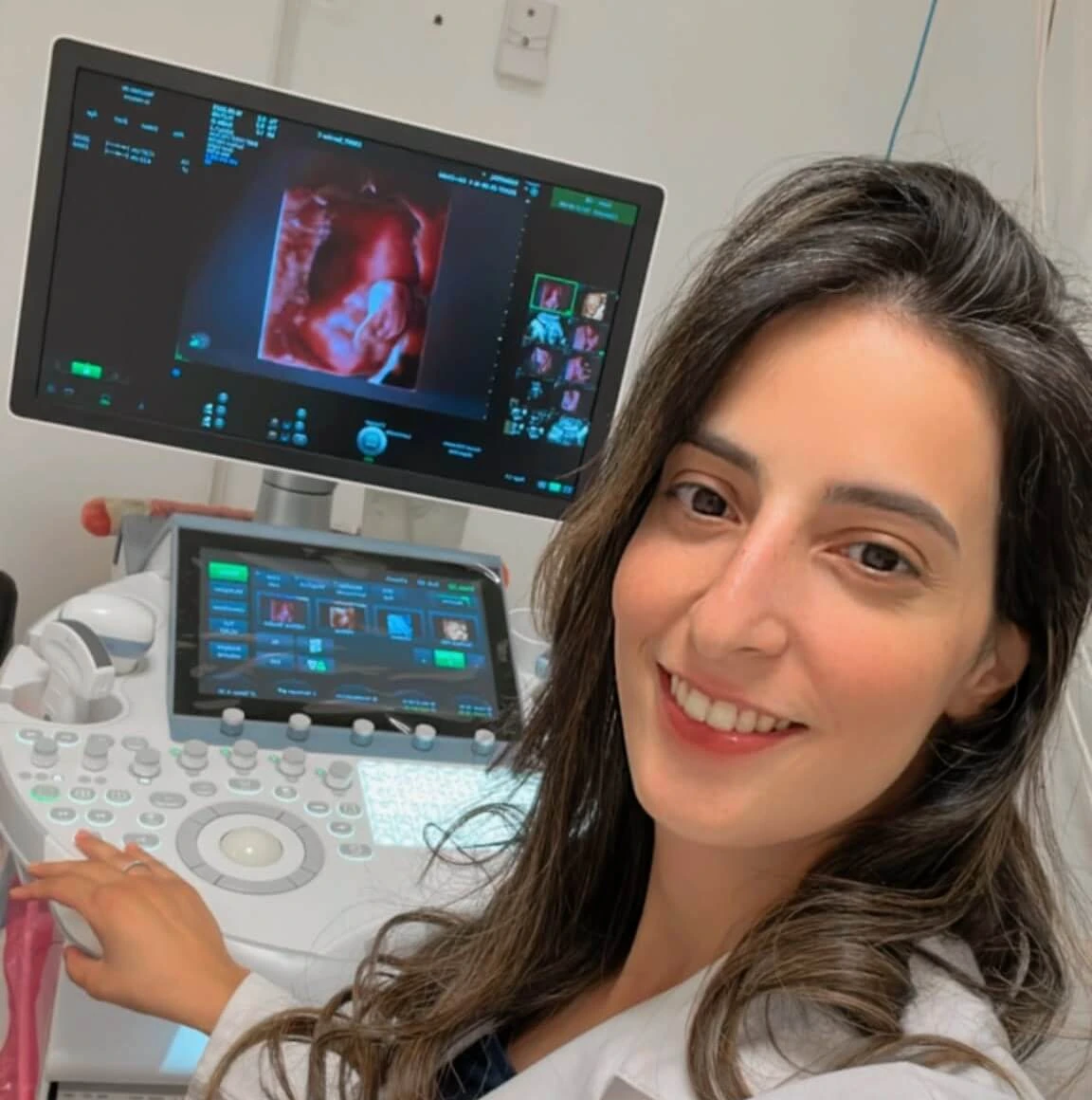
Understanding Before Interpreting
An Image Only Makes Sense in Context.
Ultrasound uses sound waves, rather than X-rays, to visualize certain parts of the body. Its purpose depends on the clinical question: dating a pregnancy, examining fetal development, assessing the uterus or exploring the ovaries.
The timing and route of the examination are part of medical decision-making. A repeat examination may be advised if imaging is incomplete or a question remains unanswered.
Pregnancy Ultrasound: Dating, Anatomy and Growth
Different stages of pregnancy raise different questions. Examinations are scheduled according to gestational age, medical history and the development of your pregnancy, rather than using the same number of scans for every patient.
Early Pregnancy and Dating
An early ultrasound may help clarify the location of the pregnancy, how it is developing and the gestational age, depending on what can be seen at that stage.
An examination performed very early may be inconclusive. Further ultrasound or laboratory testing may then be necessary, without drawing conclusions too quickly from an incomplete image.
First-Trimester Ultrasound
This examination provides information about gestational dating, the number of fetuses and certain aspects of early development. Measurements and observations are explained in their context.
Depending on the situation, the scan forms part of a broader discussion about prenatal screening. Ultrasound does not replace all screening tests and cannot exclude every abnormality.
Fetal Anatomy Scan
The anatomy scan assesses fetal structures that can be seen by ultrasound. It contributes to monitoring development and assessing growth, the placenta and amniotic fluid.
The baby’s position and imaging conditions may limit some views. A repeat scan or specialist opinion may be needed to complete the assessment.
Growth and the Third Trimester
Measurements help assess growth and estimate fetal weight. The examination may also provide information about fetal presentation, the placenta and amniotic fluid.
Estimated fetal weight remains an approximation. It is considered alongside gestational age, previous measurements and the medical context; it cannot by itself predict how birth will proceed.
Obstetric Doppler and Targeted Monitoring
Doppler ultrasound evaluates blood flow in selected vessels. It may complement monitoring when medically indicated, for example when assessing possible growth concerns.
Its usefulness depends on the vessel examined, gestational age and other results. Doppler is not necessary at every appointment and does not replace assessment of symptoms or fetal movements.
A normal ultrasound result is useful information, not a guarantee that every condition has been excluded. Some abnormalities cannot be seen, may develop later or require other investigations.
Pelvic Ultrasound: The Uterus, Endometrium and Ovaries
Gynecological ultrasound may help investigate symptoms or monitor a known condition. Images are always considered alongside clinical examination, the menstrual cycle and your medical history.
Pelvic Pain
The scan can look for certain findings that may help explain pain. Its usefulness depends on symptoms and the specific medical question.
A normal-looking ultrasound does not rule out every cause of pain. Further assessment may still be required.
Heavy Periods and Unusual Bleeding
Examination of the uterus and its lining, the endometrium, can contribute to assessment. Interpretation takes account of age, the menstrual cycle and any treatments.
Depending on the context, ultrasound may be supplemented by other tests. It does not automatically replace an indicated tissue sample or further examination.
Ovarian Cysts and Fibroids
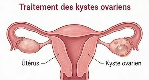

Ultrasound can describe the appearance, size and location of certain abnormalities. Monitoring or another examination may be recommended depending on what is observed.
Finding an abnormality on ultrasound does not automatically mean surgery is necessary. Decisions depend on several factors, not size alone.
Ovaries and Fertility Assessment
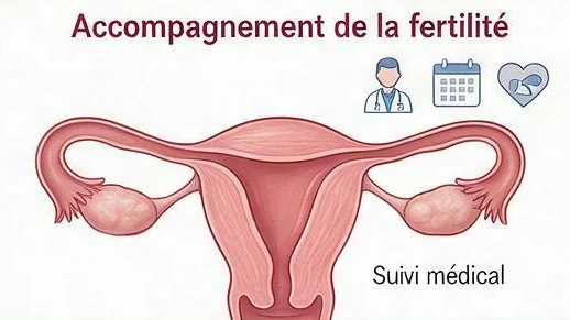
When indicated, ultrasound can contribute to assessing the ovaries, the uterine lining or cycle monitoring. The appropriate timing is discussed with the team.
Ultrasound alone does not provide a complete assessment of fertility. It is combined with other investigations and cannot guarantee a pregnancy.
Abdominal or Transvaginal Ultrasound: How Does the Examination Work?
The route of examination depends on the medical question and the imaging conditions. It is explained beforehand, respecting your consent and comfort.
Abdominal Ultrasound
A probe is moved over the lower abdomen after gel is applied. This approach produces images through the abdominal wall.
A full bladder may be requested for some pelvic examinations. You will be given preparation instructions when arranging the appointment.
Transvaginal Ultrasound
An appropriate covered and lubricated probe is introduced into the vagina to examine certain pelvic structures at closer range. This approach may provide additional information.
Your consent is required. You may decline or ask for the examination to stop. Other approaches and their possible limitations can then be discussed.
Tell the clinician about pain, anxiety, latex allergy or a difficult previous examination. You can ask what support arrangements are available.
Preparing for Your Ultrasound Appointment
-
Explain the Reason for the Scan
Say whether the examination concerns pregnancy, a gynecological symptom or a requested follow-up. Mention the known gestational age or date of your last period.
-
Confirm Preparation Instructions
Ask whether a full bladder is needed. Preparation depends on the examination route. Do not fast or undertake special preparation unless instructed.
-
Bring Relevant Records
Bring previous scans, relevant blood-test results, a referral if available and a list of medications. Comparing results can help assess change over time.
Understanding Results and Next Steps
Results are more than an image. The important questions are what the examination can show, what remains uncertain and whether further steps are needed.
Findings Explained
Measurements are interpreted in the context of the menstrual cycle, gestational age or symptoms. One number alone cannot establish a diagnosis.
A Repeat Examination When Helpful
Incomplete images, a scan performed too early or a finding needing clarification may require a repeat appointment. A specialist opinion may also be suggested.
Continuity with Your Care
Keep your reports and share them with the professionals involved in your care. If another team follows you, explain the reason for the scan to support interpretation and the next steps.
Essential Information
Your Questions About Ultrasound
Do I Need a Full Bladder?
It depends on the examination. A full bladder may help during abdominal pelvic ultrasound, whereas an empty bladder is generally requested for transvaginal scans. Follow the instructions given for your appointment.
Is Transvaginal Ultrasound Mandatory?
No. It is offered when it may be useful and requires your consent. You may refuse or ask to stop. Another approach may be discussed, including any limitations in what can be seen.
Does a Normal Ultrasound Rule Out Every Condition?
No. Ultrasound answers certain medical questions, not all of them. Some conditions cannot be seen or require other tests. Persistent symptoms should be reassessed, even after reassuring imaging.
Do 3D or 4D Images Replace a Medical Ultrasound?
No. Three-dimensional imaging does not replace a full medical examination or its measurements. If such imaging is considered, its suitability and availability should be confirmed with the practice. A picture of the baby’s face is not a comprehensive health assessment.
Can Patients Travel from Tunis or Other Regions?
The practice welcomes patients from Tunis and other regions of Tunisia by appointment. Before travelling, clarify the requested examination and which documents you have.
When a scan depends on gestational age or a particular point in the menstrual cycle, the appointment must fit the appropriate clinical schedule. Confirm this before arranging travel.
When Should I Not Wait for a Scheduled Appointment?
Severe pain, heavy bleeding, faintness or worsening symptoms require prompt medical assessment. During pregnancy, a significant reduction in fetal movements should not wait until the next ultrasound appointment.
In an emergency, seek appropriate healthcare near your location without delay. Routine practice contact channels are not an emergency service.
Our Commitment
Helping You Understand, Not Just Look at Images.
Attention to your questions, your comfort and what the results mean for you.
Ultrasound is part of a relationship of care: an appropriate indication, a consented examination and explanations of both findings and their limitations.
The Practice
Your Ultrasound Appointment in Ennasr, Ariana
Dr Ines Ben Hassen Hached welcomes you at the practice for gynecological or obstetric follow-up. When booking, explain the reason for the examination and any instructions from the referring clinician.
Located in Ennasr, Ariana Governorate, the practice welcomes patients from Tunis and other regions of Tunisia.
Dr Ines Ben Hassen Hached
First Floor, Office BA5 bisResidence La Coupole
2 Avenue Hédi Nouira, Ennasr
Ariana 2037, Tunisia
+216 27 550 875
+216 25 333 530
Consultations are by appointment. Confirm the fee, examination type and required preparation before your visit. Do not send full medical records until you have asked about an appropriate secure transmission method.
Medical Information and References
This page provides general information and does not replace a consultation. Examination schedules and arrangements mentioned in international references do not automatically apply in Tunisia. Your follow-up is adapted to your clinical situation.
- NHS — Ultrasound: Procedure and Preparation (in English)
- NHS Inform — Pregnancy Ultrasounds and the Limitations of Screening (in English)
- Guy’s and St Thomas’ NHS — Transvaginal Ultrasound (in English)
- Chelsea and Westminster Hospital — Pelvic Ultrasound (in English)
- NHS — Ultrasound scans in pregnancy